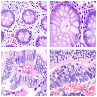
This paper examines the performance of different machine and deep learning algorithms in classifying colon histological images using different feature extraction methods. The relationship between the feature extraction methods and the selected machine learning methods to improve the classification accuracy is analyzed. Widely used methods like local binary patterns, histograms of oriented gradients, Gabor filter and Dobeshi wavelets are investigated for feature extraction from colon histological images. The features extracted by histogram of oriented gradients and Gabor filter methods were used as a single joint feature vector. And popular machine learning methods such as Support vector machine, Decision trees, Random forest, k-nearest neighbors and Naive Bayesian method were used to classify the selected images. The paper also investigates ensemble methods using gradient bousting and voting classifier as examples. The authors also focus on the study of convolutional neural networks as they are one of the main deep learning methods at the moment. The classification methods selected for analysis are compared in terms of classification accuracy and time taken for training and recognition. All pre-defined and adjustable parameters of both feature extraction methods and classification methods were personally selected by the authors as a result of experimental studies, which were conducted using a software tool created in the Python programming language on a set of LC25000 histological images. The software created is easily customizable and can be used in the future to investigate classification methods on other datasets.
| Published in | American Journal of Artificial Intelligence (Volume 8, Issue 2) |
| DOI | 10.11648/j.ajai.20240802.12 |
| Page(s) | 41-47 |
| Creative Commons |
This is an Open Access article, distributed under the terms of the Creative Commons Attribution 4.0 International License (http://creativecommons.org/licenses/by/4.0/), which permits unrestricted use, distribution and reproduction in any medium or format, provided the original work is properly cited. |
| Copyright |
Copyright © The Author(s), 2024. Published by Science Publishing Group |
LBP, HOG, Dobeshi, Feature Extraction, Classification
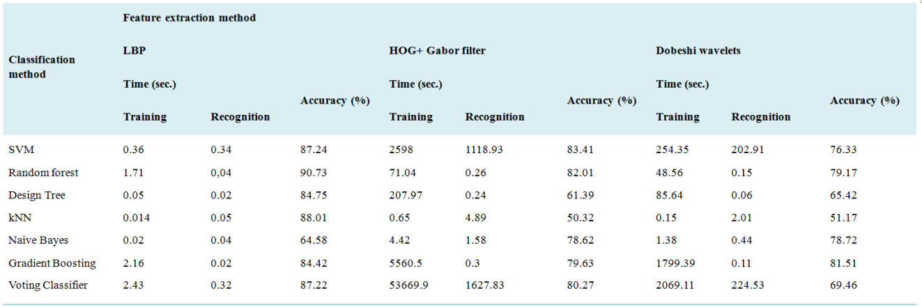
Classification method | Time (sec.) | Accuracy (%) | |
|---|---|---|---|
Training | Recognition | ||
CNN | 12936.11 | 194.7 | 98,2 |
HOG | Histograms of Oriented Gradients |
LBP | Local Binary Patterns |
CNN | Convolutional Neural Networks |
DW | Dobeshi Wavelets |
DWT | Discrete Wavelet Transforms |
SVM | Support Vector Machine |
kNN | k-Nearest Neighbor |
| [1] | Gurcan, M. N., Boucheron, L. E., Can, A., Madabhushi, A., Rajpoot, N. M. and Yener, B. Histopathological image analysis: A review. In IEEE Reviews in Biomedical Engineering, 2009, vol. 2, pp. 147-171. |
| [2] | De Matos, J., Ataky, S. T. M., de Souza Britto Jr, A., Soares de Oliveira, L. E. and Lameiras Koerich, A. Machine learning methods for histopathological image analysis: A review, Electronics. 2021, 10(5), 562. |
| [3] | Aeffner, F., Zarella, M. D., Buchbinder, N., Bui, M. M., Goodman, M. R., Hartman, D. J., Lujan, G. M., Molani, M. A., Parwani, A. V., Lillard, K. and Turner, O. C. Introduction to digital image analysis in whole-slide imaging: a white paper from the digital pathology association, Journal of pathology informatics. 2019, 10(1), p. 9. |
| [4] | Mutlag W. K. et al. Feature extraction methods: a review, Journal of Physics: Conference Series. – IOP Publishing, 2020. – Т. 1591. – №. 1. – С. 012028. |
| [5] | Dalal, N., Triggs, B. Histograms of oriented gradients for human detection. In IEEE Computer Society Conference on Computer Vision and Pattern Recognition (CVPR'05), 2005, San Diego, CA, USA, pp. 886-893 vol. 1. |
| [6] | Manjunath, B. S. and Ma, W. Y. Texture features for browsing and retrieval of image data. In IEEE Transactions on pattern analysis and machine intelligence 1996. 18(8), pp. 837-842. |
| [7] | Mallat SG. A theory for multiresolution signal decomposition: the wavelet representation. In IEEE transactions on pattern analysis and machine intelligence 1989. 11(7), pp 674-693. |
| [8] | Ojala, T., Pietikäinen, M., Harwood, D. A comparative study of texture measures with classification based on featured distributions. Pattern recognition. 1996, 29(1), pp. 51-59. |
| [9] | LeCun, Y., Bengio, Y., Hinton, G., 2015. Deep learning. Nature, 521(7553), 436-444. |
| [10] | Wyld, Henry William, and Gary Powell. Mathematical methods for physics. CRC Press, 2020. |
| [11] | Borkowski, A. A., Bui, M. M., Thomas, L. B., Wilson, C. P., DeLand, L. A. and Mastorides, S. M., 2019. Lung and colon cancer histopathological image dataset (lc25000). arXiv preprint arXiv:1912.12142. |
APA Style
Mirzaev, N., Meliev, F. (2024). Investigation of Histological Image Classification Methods Using Different Feature Extraction Techniques. American Journal of Artificial Intelligence, 8(2), 41-47. https://doi.org/10.11648/j.ajai.20240802.12
ACS Style
Mirzaev, N.; Meliev, F. Investigation of Histological Image Classification Methods Using Different Feature Extraction Techniques. Am. J. Artif. Intell. 2024, 8(2), 41-47. doi: 10.11648/j.ajai.20240802.12
AMA Style
Mirzaev N, Meliev F. Investigation of Histological Image Classification Methods Using Different Feature Extraction Techniques. Am J Artif Intell. 2024;8(2):41-47. doi: 10.11648/j.ajai.20240802.12
@article{10.11648/j.ajai.20240802.12,
  author = {Nomaz Mirzaev and Farkhod Meliev},
  title = {Investigation of Histological Image Classification Methods Using Different Feature Extraction Techniques
},
  journal = {American Journal of Artificial Intelligence},
  volume = {8},
  number = {2},
  pages = {41-47},
  doi = {10.11648/j.ajai.20240802.12},
  url = {https://doi.org/10.11648/j.ajai.20240802.12},
  eprint = {https://article.sciencepublishinggroup.com/pdf/10.11648.j.ajai.20240802.12},
  abstract = {This paper examines the performance of different machine and deep learning algorithms in classifying colon histological images using different feature extraction methods. The relationship between the feature extraction methods and the selected machine learning methods to improve the classification accuracy is analyzed. Widely used methods like local binary patterns, histograms of oriented gradients, Gabor filter and Dobeshi wavelets are investigated for feature extraction from colon histological images. The features extracted by histogram of oriented gradients and Gabor filter methods were used as a single joint feature vector. And popular machine learning methods such as Support vector machine, Decision trees, Random forest, k-nearest neighbors and Naive Bayesian method were used to classify the selected images. The paper also investigates ensemble methods using gradient bousting and voting classifier as examples. The authors also focus on the study of convolutional neural networks as they are one of the main deep learning methods at the moment. The classification methods selected for analysis are compared in terms of classification accuracy and time taken for training and recognition. All pre-defined and adjustable parameters of both feature extraction methods and classification methods were personally selected by the authors as a result of experimental studies, which were conducted using a software tool created in the Python programming language on a set of LC25000 histological images. The software created is easily customizable and can be used in the future to investigate classification methods on other datasets.
},
 year = {2024}
}
TY  - JOUR
T1  - Investigation of Histological Image Classification Methods Using Different Feature Extraction Techniques

AU  - Nomaz Mirzaev
AU  - Farkhod Meliev
Y1  - 2024/08/20
PY  - 2024
N1  - https://doi.org/10.11648/j.ajai.20240802.12
DO  - 10.11648/j.ajai.20240802.12
T2  - American Journal of Artificial Intelligence
JF  - American Journal of Artificial Intelligence
JO  - American Journal of Artificial Intelligence
SP  - 41
EP  - 47
PB  - Science Publishing Group
SN  - 2639-9733
UR  - https://doi.org/10.11648/j.ajai.20240802.12
AB  - This paper examines the performance of different machine and deep learning algorithms in classifying colon histological images using different feature extraction methods. The relationship between the feature extraction methods and the selected machine learning methods to improve the classification accuracy is analyzed. Widely used methods like local binary patterns, histograms of oriented gradients, Gabor filter and Dobeshi wavelets are investigated for feature extraction from colon histological images. The features extracted by histogram of oriented gradients and Gabor filter methods were used as a single joint feature vector. And popular machine learning methods such as Support vector machine, Decision trees, Random forest, k-nearest neighbors and Naive Bayesian method were used to classify the selected images. The paper also investigates ensemble methods using gradient bousting and voting classifier as examples. The authors also focus on the study of convolutional neural networks as they are one of the main deep learning methods at the moment. The classification methods selected for analysis are compared in terms of classification accuracy and time taken for training and recognition. All pre-defined and adjustable parameters of both feature extraction methods and classification methods were personally selected by the authors as a result of experimental studies, which were conducted using a software tool created in the Python programming language on a set of LC25000 histological images. The software created is easily customizable and can be used in the future to investigate classification methods on other datasets.

VL  - 8
IS  - 2
ER  -
Laboratory of Biometric Systems, Digital Technologies and Artificial Intelligence Research Institute, Tashkent, Uzbekistan
Biography: Mirzaev Nomaz: Since 2020 – Doctor of Technical Sciences. He defended his doctoral dissertation in 2020 at the Tashkent University of Information Technologies on the topic “Methods for the synthesis of recognition operators in the space of large characters” 01/05/03 - Theoretical foundations of computer science. From 2021 to the present, he is the project manager at the Research Institute for the Development of Digital Technologies and Artificial Intelligence. He has published 150 scientific papers, 50 of them after defending dissertations.
Research Fields: Pattern recognition, mathematical statistics, probability theory, machine learning

Laboratory of Biometric Systems, Digital Technologies and Artificial Intelligence Research Institute, Tashkent, Uzbekistan
Biography: Meliev Farkhod is a Doctoral student at the Research Institute for the Development of Digital Technologies and Artificial Intelligence. Conducts research in the field of Digital technologies and artificial intelligence. His research activities focus on the application of advanced methods and technologies for visual data analysis and processing. His work includes the study of various algorithms and approaches aimed at improving the quality and accuracy of automatic image processing. Also explores the application of popular machine learning methods in the field of medical diagnostics.
Research Fields: Image processing, machine learning